Soubor:Hematopoiesis (human) diagram.png
Plná velikost ((3 472 × 2 280 pixelů, velikost souboru: 1,18 MB, MIME typ: image/png))
Popis
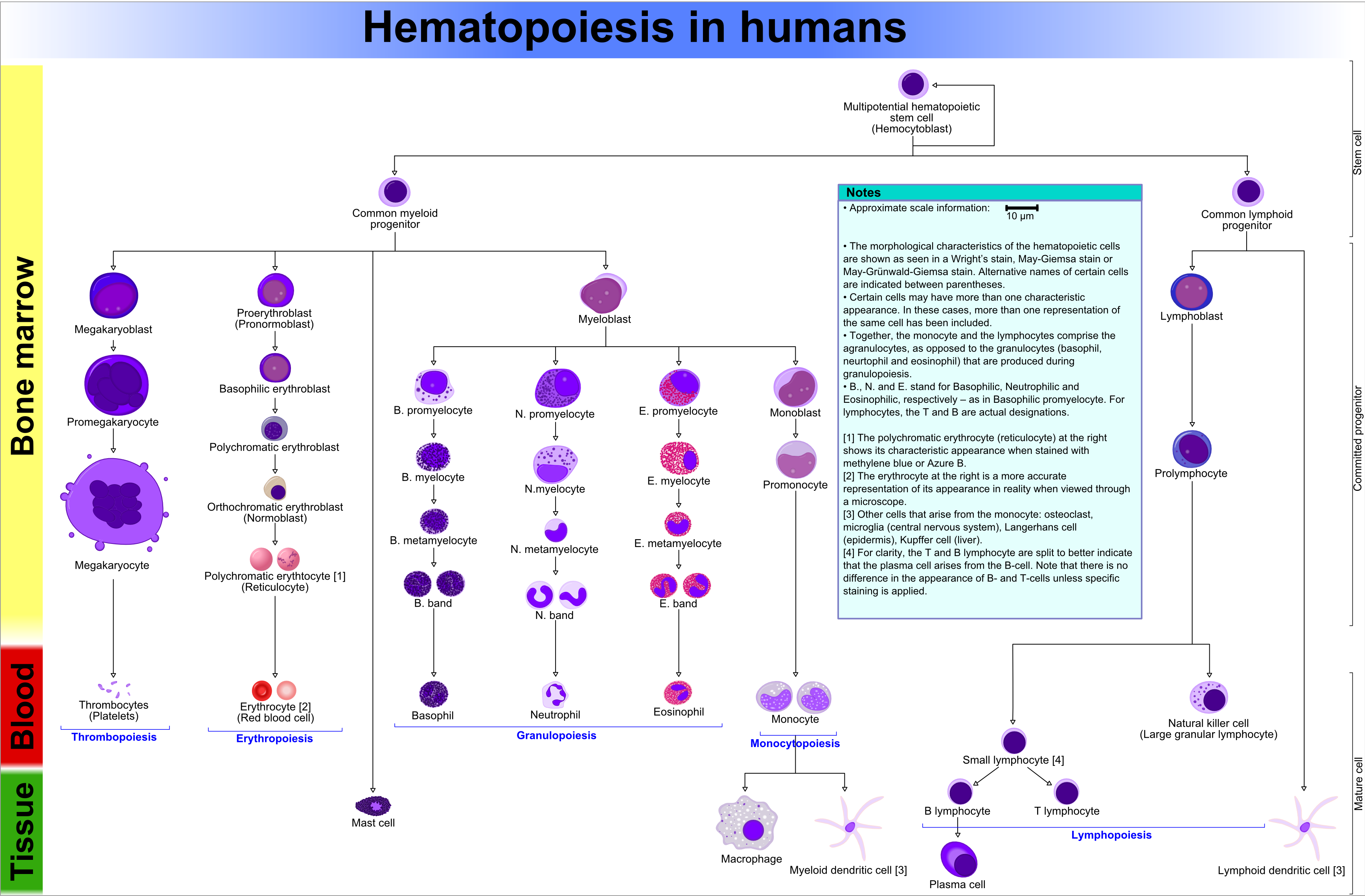
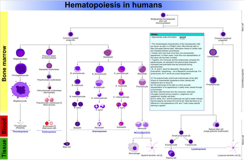
| Popis | This diagram shows the hematopoiesis as it occurs in humans. | |||
| Datum | ||||
| Zdroj | Vlastní dílo | |||
| Autor | A. Rad | |||
| Svolení (Užití tohoto souboru) |
GFDL-self. This image is released under the GFDL-self license and is considered freely distributable. This image or any reproductions/customizations thereof (or any reproductions/customizations of its reproductions/customizations, and so forth) may NOT be sold without my explicit consent. | |||
| Další verze |
[] This SVG
Other SVG
PNG
PNG with notes box
|
_diagram_switch.svg&lang=en)
_diagram_switch.svg&lang=ca)
_diagram_switch.svg&lang=es)
_diagram_switch.svg&lang=gl)
_diagram_switch.svg&lang=is)
_diagram_switch.svg&lang=pt)
_diagram_switch.svg&lang=ru)
_diagram_switch.svg&lang=ja)
_diagram_switch.svg&lang=zh-cn)
_diagram.svg)
_diagram_fr.svg)
_diagram_is.png)

_diagram_zh.png)
_diagram.png)
_diagram_en.png)
_diagram-es.png)
_diagram_uk.png)

_diagram_en.svg)
_diagram.png)
- Created using Xara X¹.
- I've drawn this schematic using the sources below as a reference. However, this does of course not mean the image contains no errors. I suggest this image be reviewed by an expert on the field of hematopoiesis. Also anyone else's comments and suggestions are welcome at my en:Wikipedia user talk page.
- I've tried to show all the characteristics of the cells as much as possible. The style in which they're drawn can be compared with how caricatures are drawn. This was not an attempt to draw the cells "photorealistically". For comparison, refer to the erythrocyte that has been drawn in 2 different ways: the style I used for all cells (left) and a different style that shows how the erythrocyte looks through a microscope (right).
- A few things I'm not sure of and that may be incorrect in the diagram:
1) the thrombocyte series can (should?) contain a metamegakaryocyte that comes between the megakaryocyte and thrombocytes. Also a micromegakaryocyte has been identified, but information on these was very rare, so I couldn't include them.
2) Images on the B/N/E Promyelocyte were rare and I'm not sure if I have drawn them correctly. Especially the E. Promyelocyte.
3) I don't know whether all the other cells from the monocytic phagocytosis system (Langerhans cell, Kupffer cell etc.) can also arise from the lymphoid DC. Currently I've also included them in the lymphoid DC.
4) I don't know whether the common myeloid/lymphoid progenitors should have nucleoli. Sources report that they're indistinguishable from small lymphocytes. But they are rapidly dividing cells and therefore (I think) should have nucleoli. Currently, I've drawn them as lymphocytes.
5) Are the common myeloid/lymphoid progenitors a part of the committed progenitor or a part of the stem cells? CLP & CMP are also called common lymphoid/myeloid stem cells, so just the word "progenitor" doesn't rule anything out here...
6) According to sources, the nucleus of a lymphoblast stains reddish-purple, while the images display a blueish-purple nucleus. These images were sometimes on the same page as the text that reported this info (reddish-purple nucleus).
7) Langerhans' cell vs. Langerhans cell. Thefreedictionary.com, On-line Medical Dictionary and Stedman's Medical Dictionary say "Langerhans cell".
Licence

|
Tento dokument smí být kopírován, šířen nebo upravován podle podmínek Svobodné licence GNU pro dokumenty verze 1.2 nebo libovolné vyšší verze publikované nadací Free Software Foundation. Dokument nemá neměnné části ani texty na předním či zadním přebalu. Kopie textu licence je k dispozici v oddíle nazvaném GNU Free Documentation License. |
| Tento soubor podléhá licenci Creative Commons Uveďte autora-Zachovejte licenci 3.0 Unported | ||
| ||
| Tato licenční šablona byla k tomuto souboru přidána v rámci změny licencování. |
References
Below are the sources I used for this diagram, for your reference only (not to clog up this page).
Books
- Parham, The immune system, 2nd ed.
- Robbins et al., Pathologic Basis of disease. 7th ed. Chapter 13, Red blood cell and bleeding disorders, page 621: figure 13-1.
PPT
- http://cpmcnet.columbia.edu/student/ssn/histology/bloodbonemarrow2004.ppt
- http://ws.westernu.edu/DO2007/studymaterial/bloodhistologylab.ppt
- http://lpc1.clpccd.cc.ca.us/lpc/jgallagher/anat1/Chapter17BloodMarieb.ppt
- http://www.usd.edu/biol/faculty/swanson/histo/Kodachromes/Lab%202.ppt
- http://wberesford.hsc.wvu.edu/marrow.ppt
- http://www2.cmu.edu.tw/~cmcmt/introduction/personal/mcshih/data/mcshih-datat06.ppt
- http://instructional1.calstatela.edu/nmcquee/Micro410/Structure%20and%20Function%20of%20Leukopoietic%20Tissue.ppt
- http://meds.queensu.ca/medicine/deptmed/hemonc/dload/concepts.ppt
- http://www.leukine.com/healthcare/cascade.pdf
- http://www.iis.fraunhofer.de/medtech/med_bild/hemacam/differential_blutbild_BMT2004.pdf
- http://www.montana.edu/wwwmb/coursehome/mb405/PDF%20Files/HEMATO23.pdf
- http://www.uzleuven.be/uzroot/hosting/labo/Leermodule/GENERAL_LAB_MED/HEMATOLOGY/Documenten/LES_Leucopoiese_Afwijkingen_Leukocyten.pdf
- http://intl.elsevierhealth.com/e-books/pdf/54.pdf
- http://bio-bg.net/materials/kuby_immunology/Kuby%20Immunology/Chapter%2002.pdf
Websites
- [1] - Schematic with info on blood cells
- [2] - info on blood cells and their progenitors.
- [3] - More info on blood cells (erythroid series amongst others).
- PMID 15123777 →Figure 6
- PMID 16455345
- [4] - Hematopoiesis schematic
- [5] - Number of nucleoli of 2 hematopoietic stem cells
- [6] - Explanation of the "committed progenitor"
- [7] - characteristics of the lympho series
- [8]
- [9] - Cells from the monocytic series
- [10] - Pics of many blood cells
- [11] - Monocytic series characteristics
- [12] - Lots of hematology images
- [13] - Some progenitors of megakaryocyte (pics)
- [14] - Schematic of the myeloid and lymphoid hematopoietic system
- [15] - megakaryo progenitor slides
- [16] - Stages of maturation of megakaryo progenitors with pics
- [17] - Stages of maturation of megakaryo progenitors
- [18] archive copy at the Wayback Machine - On reticulocytes: The cytoplasmic color at this stage is called polychromatic or polychromatophilic when viewed with Wright’s stain. If stained with new methylene blue, the remaining RNA will precipitate resulting in a reticular appearance.
- [19] - On reticulocyte: Cytoplasm stains slightly basophilic with Wright's stain. However, when stained with a supravital-stain such as new methylene blue or brilliant cresyl blue, precipitated ribrosomal RNA (reticulum) can be demonstrated within the cell
- [20] - Contains an index of (amongst others) hematopoietic cells with frequently used synonyms. Search ndif.org with google
- [21] - Contains hematopoietical cellular characteristics and synonyms of all the progenitors
- [22] - Contains sizes in microns of granulo's + progenitors and cells in erythroid series and other hematopoietic cells (agranulo's)
- [23] - index of histology pictures. White and red blood cells and progenitors included
- [24] - Some basic info on hematopoiesis. Sizes of erthroid progenitors are given too
- [25] - Contains erythroid cells in the order of maturation, categorized per granulocyte. Staining info included
- [26] - Easy to understand overview of hematopoiesis. Erythroid and myeloid series discussed. Also thrombopoiesis is discussed
- [27] - Contains ASCII schematics on hematopoiesis, and on the growth factors, CFU notation and end cells
- [28] - Contains granulocytic progenitor cells. Band form of eo and baso missing
- [29] - Hematopoietic system. Figure is pretty complete and contains good representations of the progenitor cells. Found by typing the following in google: eosinophil metamyelocyte
- [30] - Good tute on Hematopoiesis. Schematic pics are a bit ugly, but the general picture is clear
- [31] - Contains myeloid cells (granulo's) in the order of maturation, categorized per granulocyte. Staining info included
- [32] - hematopoiesis pics in Mr.Sid format (.sid). Browser plugin for viewing the pics required
- [33] - American Society of Hematology contains hematological pics in online publication (ePub) format.
- [34] - Hematology pics of different cell series (ERYTHROPOIESIS GRANULOPOIESIS MONOCYTIC SERIE LYMPHOPOIESIS THROMBOPOIESIS CYTOCHEMICAL STAINS)
- [35] - Hematology images with short explanations. Not all progenitors are included
- [36] - Hematology atlas with many images on different progenitors of cells. Many different categories
- [37] - Hematology pics with the maturation sequence (progenitors & mature cells) and some pathologic pics
- [38] - Slide on the basophilic myelocyte + characteristics. Author discusses how the basophilic myelocyte is not discernible from the basophilic PROmyelocyte
- [39] - Some info on how the progenitor cells in hemopoiesis look (characteristics)
- [40] - Extended info on hematopoiesis with a diagram and some tables with normal values of frequencies of hematopoietic cells
- [41] - Contains erythroid progenitor names ? Proerythroblast ? Basophilic Erythroblast ? Polychromatophilic Erythroblast ? Normoblast (orthochromatic erythroblasts) ? Reticulocyte (polychromatophilic erythrocyte) ? Mature Erythrocyte
![[1]](http://www.mybloodyourblood.org/doc/poster_bloodfacts.jpg)
Historie souboru
Kliknutím na datum a čas zobrazíte příslušnou verzi souboru.
| Datum a čas | Náhled | Rozměry | Uživatel | Shrnutí | |
|---|---|---|---|---|---|
| aktuální | 19. 3. 2016, 19:02 | | 3 472 × 2 280 (1,18 MB) | wikimediacommons>Mikael Häggström | +Common names: Platelets, RBCs |
Využití souboru
Tento soubor používají následující 3 stránky:
_diagram.png)