Soubor:Gray491.png
Gray491.png ((500 × 438 pixelů, velikost souboru: 63 KB, MIME typ: image/png))
Popis
| Popis |
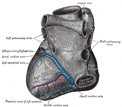
Deutsch: Blick von hinten auf das Herz. Darstellung von Henry Gray. |
||||||||||||||||||||
| Plate | 491 | ||||||||||||||||||||
| Datum | před 1858 | ||||||||||||||||||||
| Zdroj |
|
||||||||||||||||||||
| Autor |
|
||||||||||||||||||||
.jpg)
Kniha
| Henry Gray: Gray's Anatomy (20. vydání)
|
|||||||||||||||||||||||
|---|---|---|---|---|---|---|---|---|---|---|---|---|---|---|---|---|---|---|---|---|---|---|---|
| Autor |
|
-_Title_page.png) | |||||||||||||||||||||
| Editor |
Revised by Warren H. Lewis |
||||||||||||||||||||||
| Ilustrátor |
|
||||||||||||||||||||||
| Název | |||||||||||||||||||||||
| Edice |
20 |
||||||||||||||||||||||
| Vydavatel | |||||||||||||||||||||||
| Druh objektu |
verze, vydání nebo překlad |
||||||||||||||||||||||
| Seznam stran | list of all the plates | ||||||||||||||||||||||
| Jazyk |
angličtina |
||||||||||||||||||||||
| Datum vydání |
1918 |
||||||||||||||||||||||
| Místo vydání |
Filadelfie / New York |
||||||||||||||||||||||
| Zdroj | Bartleby | ||||||||||||||||||||||

Licence
Tento obrázek je volným dílem, protože se jedná jen o mechanický sken či fotokopii originálu volného díla anebo se jedná o dílo, jež se evidentně takovému skenu či fotokopii podobá natolik, že nemůže požívat ochrany autorského práva. Originál samotný je volným dílem z následujícího důvodu:
This tag is designed for use where there may be a need to assert that any enhancements (eg brightness, contrast, colour-matching, sharpening) are in themselves insufficiently creative to generate a new copyright. It can be used where it is unknown whether any enhancements have been made, as well as when the enhancements are clear but insufficient. For known raw unenhanced scans you can use an appropriate {{PD-old}} tag instead. For usage, see Commons:When to use the PD-scan tag.  | ||||
The coronary sinus is a collection of veins joined together to form a large vessel that collects blood from the myocardium of the heart. It is present in humans and other animals. It delivers deoxygenated blood to the Right atrium in conjunction with the superior and inferior vena cava.
The coronary sinus opens into the right atrium, between the inferior vena cava and the atrio-ventricular orifice. It returns the blood from the substance of the heart, and is protected by a semicircular fold of the lining membrane of the auricle, the coronary valve (the valve of Thebesius). The sinus, before entering the auricle, is considerably dilated - nearly to the size of the end of the little finger. Its wall is partly muscular, and at its junction with the great coronary vein is somewhat constricted and furnished with a valve consisting of two unequal segments.(Gray 462)
Location: It is located in the right atrium and runs transversely in the groove between the left atrium and ventricle on the posterior surface of the heart.
The coronary sinus orifice (opening) is just superior to the septal leaflet of the tricuspid valve. The coronary sinus orifice is also known as the ostium of the coronary sinus, and is guarded by the Thebesian valve.
Drainage: It receives blood mainly from the small, middle, great and oblique cardiac veins. It also receives blood from the left marginal vein and the left posterior ventricular vein. The anterior cardiac veins drain directly into the right atrium. (Some small veins drain into any of the four chambers of the heart.)
It drains into the right atrium on the posterior, inferior surface, medial to the inferior vena cava opening.
Historie souboru
Kliknutím na datum a čas zobrazíte příslušnou verzi souboru.
| Datum a čas | Náhled | Rozměry | Uživatel | Shrnutí | |
|---|---|---|---|---|---|
| aktuální | 23. 1. 2007, 22:35 | | 500 × 438 (63 KB) | wikimediacommons>Pngbot | optimized with optipng |
Využití souboru
Tento soubor používají následující 2 stránky: